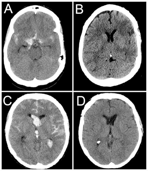
Fig. 3.

Computed tomography scans obtained in representative patients from the control and heparin groups. A and B: Admission (A) and follow-up (B) CT scans obtained in a 50-year-old patient who presented with a ruptured anterior communicating artery aneurysm. The patient was neurologically intact upon presentation. The patient underwent successful surgical clipping. The patient’s hospital course was complicated by clinical vasospasm with associated aphasia. Despite multiple intravascular interventions, the patient developed frontal and parietal infarcts. At follow-up, the patient’s aphasia was improving. C and D: Admission (C) and follow-up (D) CT scans obtained in a 47-year-old patient who presented with a ruptured anterior communicating artery aneurysm. The patient was comatose upon presentation with a GCS score of 6T. The patient underwent successful surgical clipping and was subsequently treated with a low-dose intravenous heparin infusion. The patient developed angiographic vasospasm but did not require rescue intervention. At follow-up, the patient was neurologically normal, including normal cognition and short-term memory.
