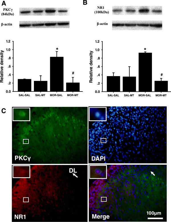
Figure 3.

Effects of melatonin on morphine-induced increased expression of PKCγ and NR1 in the spinal cord. Western blot shows expression of PKCγ (A) and NR1 (B) in the spinal cord dorsal horn (n = 3) in each sample following treatment of morphine with or without melatonin for consecutive 14 days. Data are presented as mean ± SEM. *P < 0.05, compared with SAL-SAL group # P < 0.05, compared with MOR-SAL group. C: Co-localization of spinal PKCγ and NR1. There was co-localization of PKCγ and NR1 immunoreactivity in the superficial layers (I and II) of the spinal cord dorsal horns at the lumbar (L4) level. Spinal cord samples were taken from rats receiving a combination of morphine and melatonin for consecutive 14 days (n = 3). Blue: DAPI for nucleus. Scale bar: 100 μm. DL: the dorsolateral part of the spinal cord dorsal horn.
