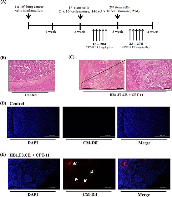
Figure 5. Histopathological analysis and tumor-tropic effect of stem cells in metastatic lung cancer animal models.

A549 lung cancer cells (1 × 10⁵ cells/mouse) were implanted in the right hemisphere of mice and CM-DiI pre-stained stem cells (1 × 10⁵ cells/mouse) were injected into the left hemisphere two times. To induce the therapeutic effect of stem cells, CPT-11 (13.5 mg/kg/day) was administered by intraperitoneal injection (i.p.) for five days. After final prodrug injection, all of the mouse brain was excised and the specimen was subjected to hematoxylin and eosin (H&E) staining for histological analysis. (A) Scheme of metastatic lung cancer animal models. (B) Negative control brain specimen. (C) HB1.F3.CE and CPT-11 co-treated brain specimen. (D) Negative control group. The in vivo migratory ability was determined by fluorescence analysis through DAPI staining of brain specimens. (E) HB1.F3.CE and CPT-11 treated group. Blue fluorescence: DAPI stained nucleus of A549 lung cancer cells and HB1.F3.CE cells. White arrow: migrated stem cells. Magnification × 100 and × 200.
