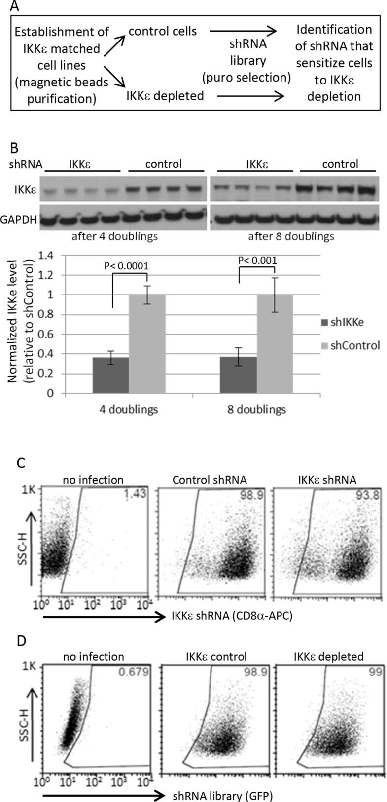
Figure 1. Human kinome shRNA screening.

(A) Dual shRNA screening procedure (B) The magnetic beads-based purified cells followed by shRNA library infection were maintained for indicated times upon completion of puromycin selection for 4 days, and then harvested for Western blotting in four biological replicates. For quantification, the signals were quantified by ImageJ software. IKKε expression was normalized by GAPDH level. The statistical significance was determined by 2-sided t-test. (C) The purity of Ovcar5 CD8α-positive cells (IKKε-matched cell line pairs) was measured by FACS at 13 days after beads purification. (D) The shRNA library vectors co-expressing GFP were introduced into the pseudo-isogenic IKKε-control and -depleted cell line pair. The purity of shRNA library in Ovcar5 cells was measured by GFP expression 2 days after completion of puromycin selection.
