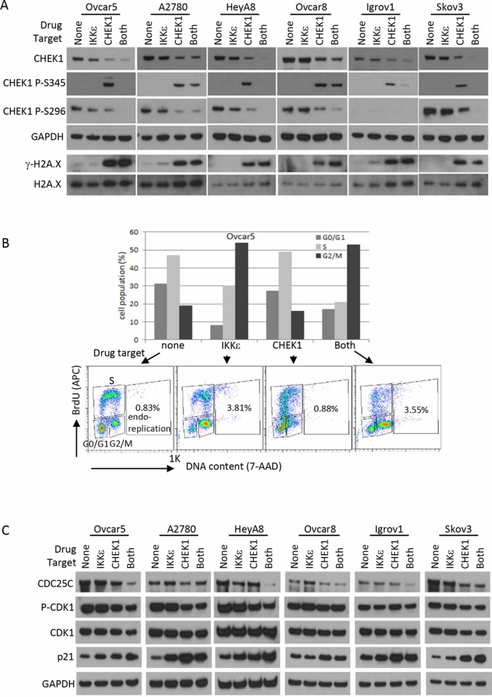
Figure 5. DNA damage response and cell cycle analysis after CHEK1 and IKKε inhibition.
(A) Western blotting for DNA damage response. Phosphorylated CHEK1, total CHEK1, and γ-H2A.X were measured by Western blotting after 24 hour treatment at final concentrations of 2 μM (BX795) and/or 0.5 μM (PF477736) prepared in fresh medium. GAPDH and H2A.X were used as loading controls. (B) Cell cycle was analyzed after pharmacological intervention in Ovcar5. Cells were treated for 16 hours at final concentrations of 2 μM (BX795) and/or 0.5 μM (PF477736) prepared in fresh medium. G0/G1, S, and G2/M phases were measured based on staining of APC-BrdU and 7-AAD by flow cytometry. (C) Western blotting for cell cycle regulators. The treatment conditions and total cellular lysates preparations are same as in Figure 4E–F.

