
Figure 6. Phagocytosis of E. coli K1 by macrophages (A–D) and microglial cells (E–H) from young and aged mice in vitro.
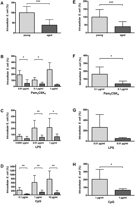
In non-activated state, phagocytosis of E. coli was significantly lower in aged compared to young macrophages (n = 9/9) (A) and in aged compared to young microglial cells (n = 15/13) (E). Macrophages from aged mice phagocytosed significantly less E. coli than macrophages from young mice after treatment with different concentrations of Pam3CSK4 (B), LPS (C), and CpG (D) (n = 6–9 per group). Similarly, microglial cells from aged mice phagocytosed less E. coli than microglial cells from young mice after treatment with a selected dose of Pam3CSK4 (F), LPS (G), and CpG (H) (n = 6 per group). Data are shown as means ± SD; *p < 0.05, **p < 0.01, ***p < 0.001, Student's t-test, Bonferroni correction in B, C, and D.