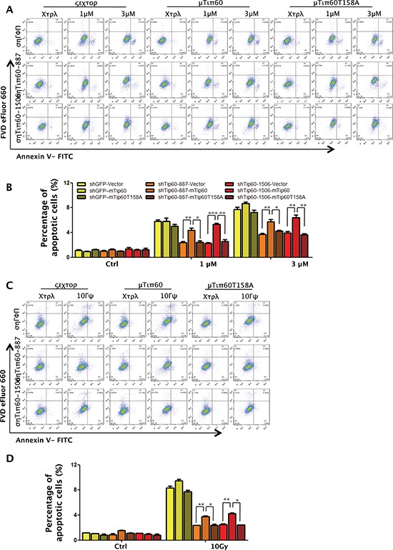
Figure 11. Phosphorylation of Tip60 at T158 by p38α is required for apoptosis induction in response to DNA damage.

(A and C) U2OS cells were co-transduced with shRNA for GFP (shGFP) or Tip60 (shTip60-887 or -1506) and vector (WN), wild type mouse Tip60 (WT) or mutant mouse Tip60 (T158A), and treated with 1 μM or 3 μM of Dox for 24 h (A) or 10 Gy of γ-radiation followed by incubation for 24 h (C). Cells were collected, stained with a FITC-conjugated anti-Annexin-V antibody and FVD eFlour 660, and analyzed by FACS. (B and D) Quantification and statistical analysis of the data in A (B) or C (D). The percentage of apoptotic cells was quantified as the percentage of FITC-positive cells in the gated area. Values are mean ± SEM for triplicates.
