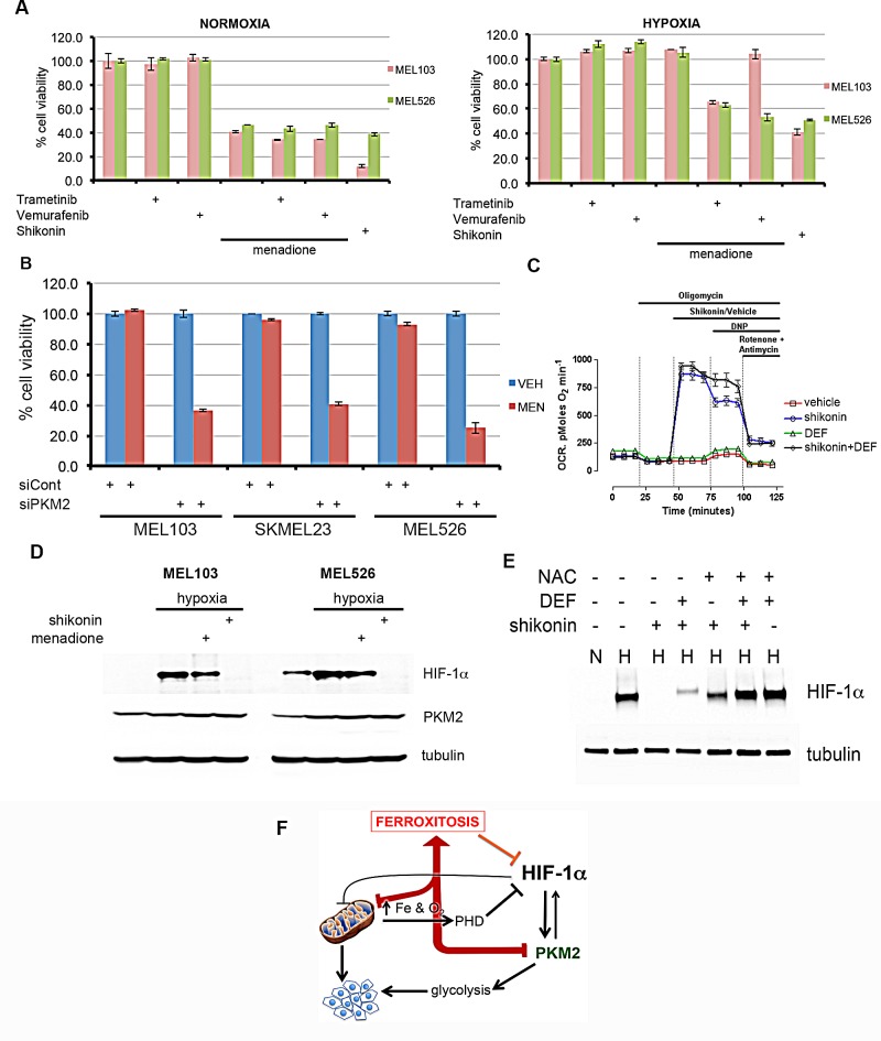
Figure 5. Activation of ferroxitosis involves concurrent inhibition of oxidative phosphorylation and PKM2-dependent glycolysis in melanoma.
A) MEL526 (BRAFV600E) or MEL103 (NRASQ61L) cells exposed to MEK inhibitor trametinib, BRAF inhibitor vemurafenib, menadione, shikonin, or a combination of these drugs as shown in normoxia or hypoxia, and cell viability measured. B) Indicated melanoma cells were transfected with control or PKM2 siRNA; treated with vehicle (blue) or MEN (red) in hypoxic conditions and cell viability assessed. C) OCR measurements of melanoma cells exposed to vehicle (DMSO) or shikonin, shikonin+DEF. Each data point represents mean OCR ± s.e. from 5 replicates. D) Western blot analysis of melanoma cells treated with menadione or shikonin under hypoxic conditions by blotting with HIF-1α or tubulin antibodies. E) MEL103 cell extracts from normoxia (N) or hypoxia (H) exposed to shikonin, N-acetyl cysteine (NAC), deferoxamine (DEF) or in combination as shown, and blotted with HIF-1α antibody. F) Activation of ferroxitosis from concurrent inhibition of oxidative phosphorylation and PKM2-dependent glycolysis.

