
Figure 1. Expression of Aurora-A protein in HCC tissues and its correlation with prognosis of patients.
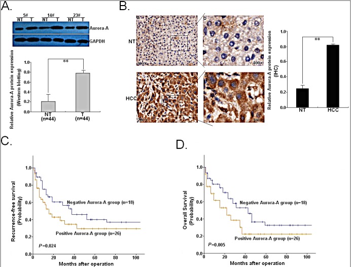
(A) Western blotting was performed to detect Aurora-A protein level in 44 paired of HCC and NTs. Three pairs of human HCC samples were exhibited. GAPDH was used as an internal control. (B) Immunohistochemical analysis. Intensive staining was observed in the cytoplasma of HCC cells, whereas less staining was showed in NTs (original magnification a, c: ×200; b, d: ×400). (C) The Kaplan-Meier survival curve of recurrence-free survival (RFS) according to Aurora-A immunostaining in 44 cases of HCC patients. (D) The Kaplan-Meier survival curve of overall survival (OS) according to Aurora-A immunostaining in 44 cases of HCC patients. Data were presented as mean ± SD of at least three independent experiments. N.S, P>0.05; *P<0.05; **P<0.01.