
Figure 6. Effects of Aurora-A upregulation on in vivo chemosensitivity of HCC cells.
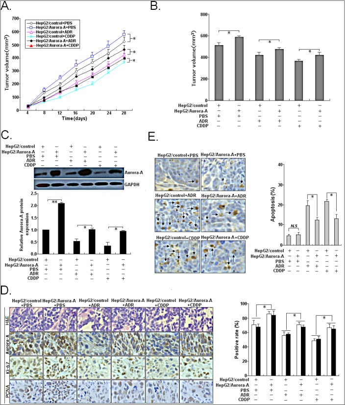
Mice were treated with ADR (2.0 mg/kg body weight; i.p., thrice), DDP (3.0 mg/kg body weight; i.p., thrice), or with 0.1ml PBS (pH 7.4; i.p., thrice). (A) Growth of tumors in the mice injected with HepG2/Aurora-A or HepG2/control cells treated with ADR, DDP or PBS. The inoculation was done in eight mice. (B) Representative features of tumors 28d after inoculation using SMMC-7721/shAurora-A or SMMC-7721/shcontrol cells treated with ADR, DDP or PBS. (C) Western blotting detection of Aurora-A protein expression in tumors developed from HepG2/Aurora-A or HepG2/control cells treated with ADR, DDP or PBS, respectively. GAPDH was used as an internal control. (D) Immunostaining of Aurora-A, Ki-67 and PCNA protein expression in tumors developed from HepG2/Aurora-A or HepG2/control cells treated with ADR, DDP or PBS. Upper: H&E staining; Intermediate and lower: immunostaining; Bars, 100μm. (E) TUNEL assay detection of apoptosis in tumors developed from HepG2/Aurora-A or HepG2/control cells treated with ADR, DDP or PBS, respectively. Data were presented as mean ± SD of at least three independent experiments. N.S, P>0.05; *P<0.05; **P<0.01.