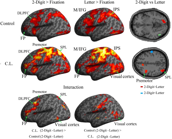
Figure 3.
Brain regions (left hemisphere) recruited in the recall phase (p < 0.001, uncorrected, k >100) were displayed on a standard inflated brain. The direct comparisons between 2-digit and letter conditions of C.L. as well as the controls were displayed on the right panel (axial plane). The FP, DLPFC, premotor cortex, and SPL were more engaged in the 2-digit condition than in the letter condition of C.L. The interactions were displayed at the bottom panel. Abbreviations: FP, frontal pole; DLPFC, dorsolateral prefrontal cortex; SPL, superior parietal lobule; IPS, intraparietal sulcus; M/IFG, middle/inferior frontal gyri.

