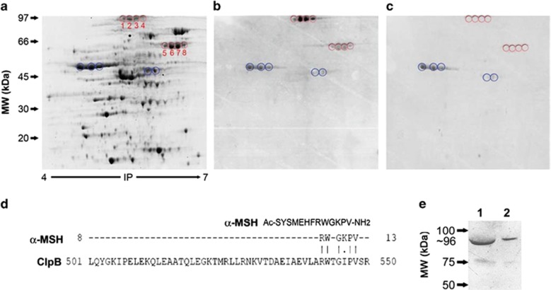
Figure 1.
Proteomic identification of molecular mimicry between E. coli K12 proteins and α-MSH. (a) 2D GE of E. coli cytoplasmic proteins. (b, c) Immunoblots of E. coli proteins detected with rabbit anti-α-MSH IgG, preadsorbed (c) or not (b) with α-MSH. Circles in red surround the spots specifically recognized by α-MSH IgG which were used for protein identification. Circles in blue indicate nonspecific spots. Proteins identified in the spots 1–4 are isoforms of ClpB. (d) α-MSH and ClpB amino-acid sequence alignments using the Stretcher program. (e) Western blot of the recombinant ClpB, revealed with anti-α-MSH IgG. Lanes 1 and 2, 20 and 10 μg of ClpB, respectively. IP, isoelectric point.

