Figure 3.

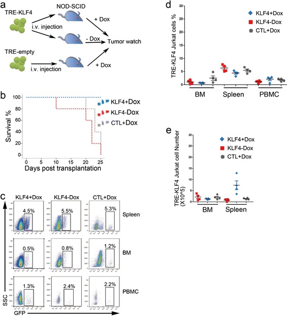
Overexpression of KLF4 in Jurkat cells in vivo. (a) Experimental design for studying KLF4 function in Jurkat cells in vivo. TRE-KLF4 Jurkat cells in which KLF4 expression was induced by Dox treatment were intravenously injected into NOD-SCID mice. The injected mice were separated into two groups (n = 15) with Dox treatment or without Dox treatment. In the third group, NOD-SCID mice were injected with TRE-empty Jurkat cells and were subsequently treated with Dox. The three groups of mice were monitored for tumors. Dox was intraperitoneally administered every two days. (b) Survival curves for the NOD/SCID mice injected with TRE-KLF4 Jurkat cells. 15 mice were used in each group. Red dots represent Dox-treated mice with injection of TRE-KLF4 Jurkat cells (KLF4 + Dox); Blue dots represent mice injected with TRE-KLF4 cells without Dox treatment (KLF4-Dox); Green dots represent Dox-treated mice with injection of TRE-empty cells (CTL + Dox). (c) Two weeks after injection of Jurkat cells, four mice from each group were culled for detection of Jurkat cells. Representative FACS profiles of mononuclear cells of spleen, BM, and peripheral blood from the three groups of mice described in b. (d) Summary of percentages of TRE-KLF4 Jurkat cells in spleen, BM, and peripheral blood from the three groups of mice described in b. (e) Summary of absolute numbers of TRE-KLF4 Jurkat cells in spleen and BM from the three groups of mice described in b.
