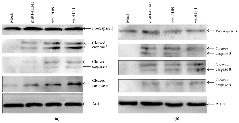
Figure 5.
Activation of apoptotic proteins in H1N1-infected HEK293 and HBE cells. Cell lysates were prepared from miRT-H1N1, scbl-H1N1, or wt H1N1-infected and uninfected HBE (a) and HEK293 (b) cells at 24 h after infection. The cell lysates were resolved with 10–15% SDS-PAGE. Proteins were transferred to PVDF membranes for western blot analyses. Analyses were performed at least three times for each protein. Cleavage of caspases 3, 8, and 9 was shown.

