
Fig. 2.
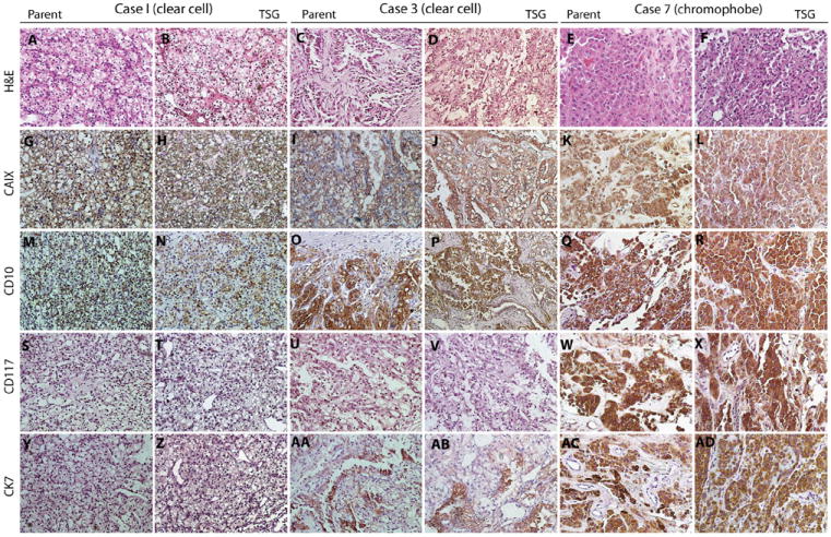
TSGs preserved histologic features and biomarker expression of corresponding parental tumors. A, C, and E are H&E-stained tumor sections from cases 1, 3, and 7, which showed similar histology to the derivative TSGs in B, D, and F, respectively. All 3 cases and their TSGs displayed expression of CAIX (G–L) and CD10 (M–R). Cases 1 and 3 and their TSGs were negative for CD117 (S–V), whereas case 7 and its TSG were positive (W and X). Case 1 and its TSG were negative for CK7 (Y and Z), and cases 3 and 7 and their TSGs were positive (AA–AD). (Color version of the figure is available online.)