
Fig. 3.
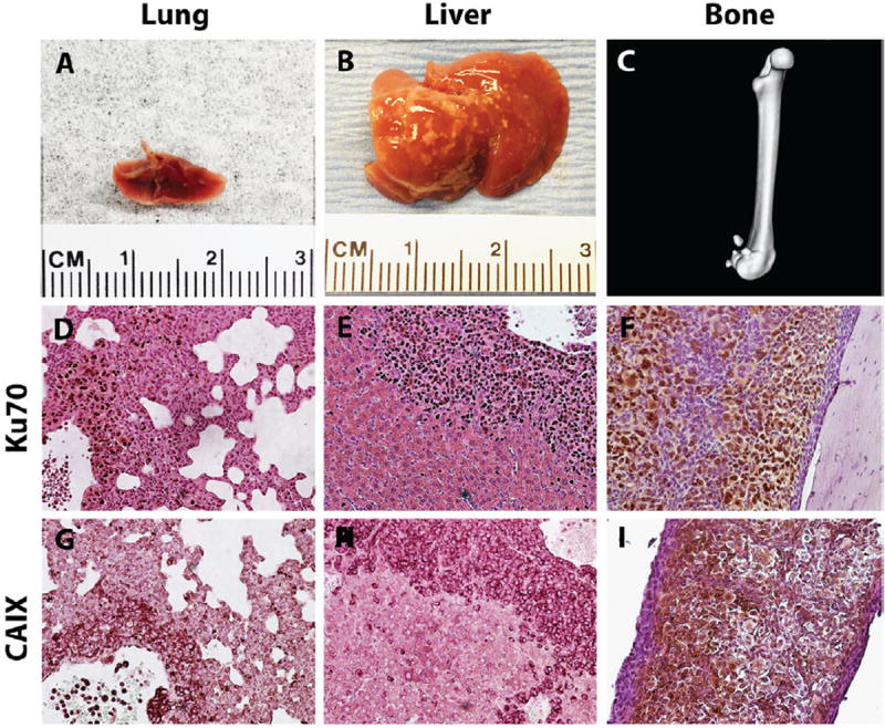
TSGs displayed metastatic potential. Gross metastases were detected in lung, A, liver, B, and bone, C, of mice bearing TSGs derived from case 1. RCC cells were distinguished from host cells at the metastatic sites by intense staining of human-specific nuclear antigen Ku70 (D–F). These cells were also positive for CAIX (G–I). (Color version of the figure is available online.)