
Fig. 1. Blood-brain barrier breakdown in the hippocampus in the living human brain during normal aging.
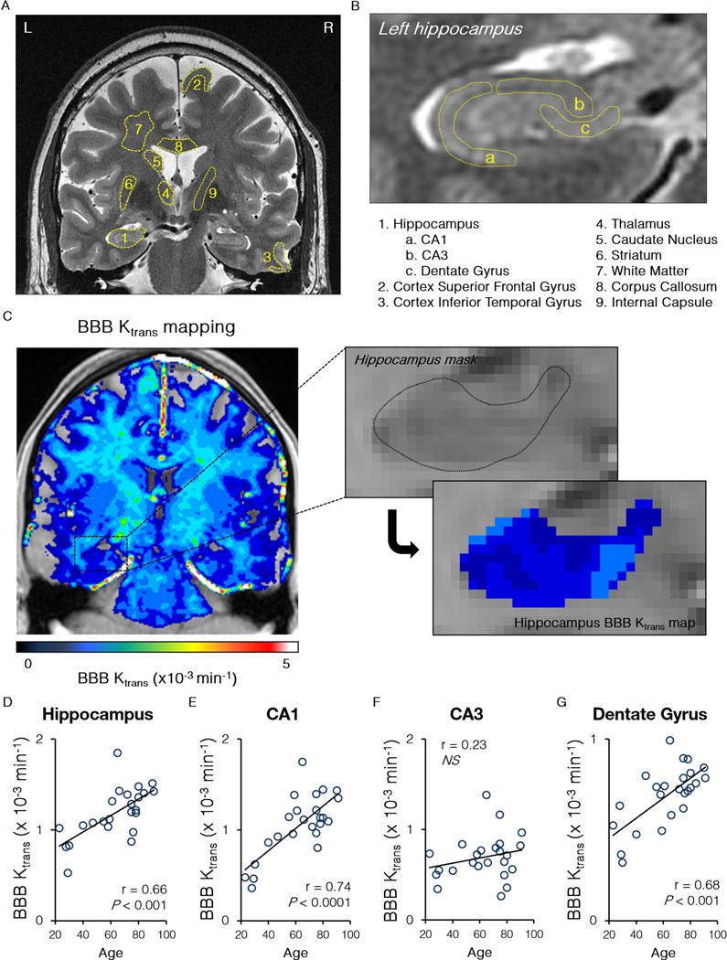
(A–B) A dynamic contrast enhanced magnetic resonance imaging was used to quantify the blood-brain barrier (BBB) regional permeability Ktrans constant in 12 regions-of-interest in the gray and white matter in the living human brain (A) including subdivisions of the hippocampus (B). (C) Representative BBB Ktrans maps of the whole brain (left) and the hippocampus (right) in a young participant with no cognitive impairment. (D–G) Age-dependent increase in the BBB permeability Ktrans constant in the entire hippocampus, its CA1 region and dentate gyrus, but not CA3 region. Single data points for the Ktrans constant from 24 individuals with no cognitive impairment (both genders, ages 23–91; see Table 1) were plotted against age; r = Pearson’s coefficient; P, significance; NS, non-significant. See also Supplementary Figure S1.