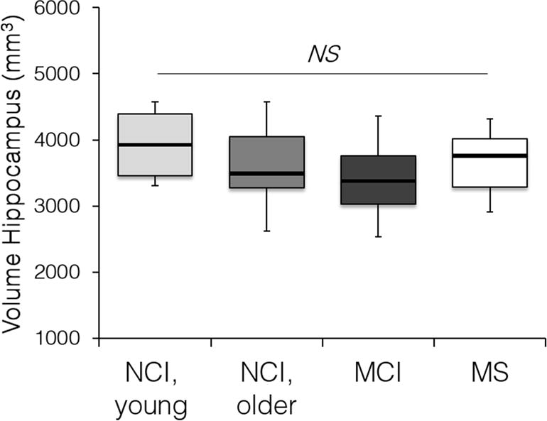
Fig. 3. Hippocampus volume in the studied groups.

Hippocampus volume was determined on T2-weighted images in individuals with no cognitive impairment (NCI), with mild cognitive impairment (MCI), and multiple sclerosis (MS) cases with no cognitive impairment. NS, non-significant by ANOVA followed by Tukey’s post hoc tests. NCI, young (n=6, ages 23–47, both genders); NCI older (n=18, ages 55–91, both genders); MCI (n=20, ages 55–85, both genders); MS (n=19, ages 26–53, both genders). See Table 1.
