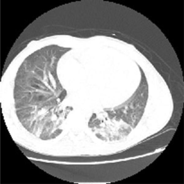
Fig. 1.
Computed tomography scan of the chest with contrast from the second day of admission showing bibasilar bronchocentric consolidative opacities with diffuse ground glass concerning for bronchopulmonary pneumonia or acute chest syndrome and a prominent main pulmonary artery which may be seen with sickle cell disease secondary to high flow state

