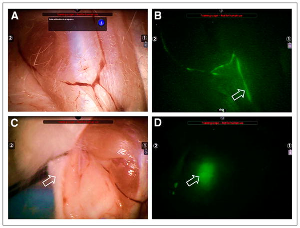
FIGURE 1.
The FireFly endoscope attached to Da Vinci Si robotic surgical system visualized all popliteal lymph nodes at 1 and 36 h after administration of 1.7 or 8.4 nmol of 99mTc-labeled fluorescent tilmanocept. (A) Bright-field image of rabbit hind-limb was acquired 1 h after 1.7-nmol injection of 800CW-tilmanocept. (B) After switch to fluorescence mode, lymphatic channel (arrow) was visualized. (C and D) On repositioning of camera to visualize popliteal region (arrow, C), fluorescence mode visualized SLN (arrow, D) without surgical exposure. SLN accumulated 0.87% of injected dose (25 pmol of IRDye800CW).

