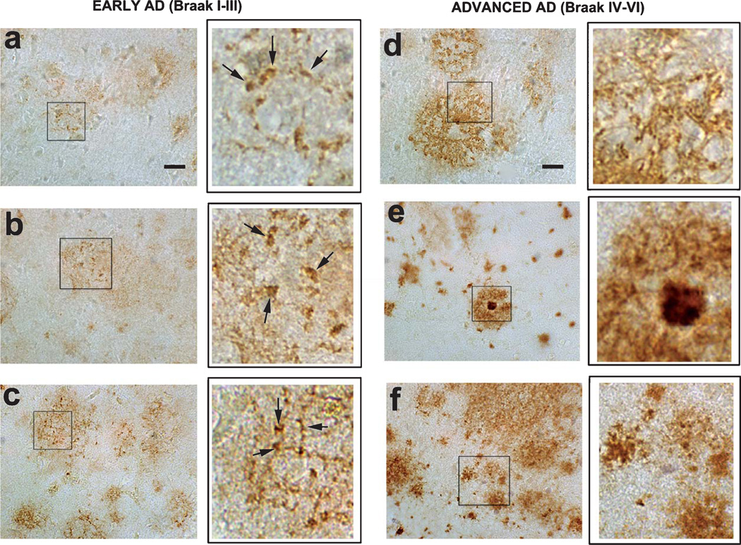
Fig. 4.
Immunohistochemical analysis of the distribution of pE(3)Aβ with the D129 antibody in early and advanced AD cases. Immunohistochemistry was conducted with the D129 antibodies in order to examine the expression patterns of pE(3)Aβ in the frontal cortex of AD at varying stages of the disease. (a–c, insets at higher magnification, arrows indicate D129 immunodense regions) representative micrographs of D129 immunoreactivity in the frontal cortex of patients with early AD (Braak stages I–III) showing differential reticular and granular region in the neuropil. (d–f, insets at higher magnification) representative micrographs of D129 immunoreactivity in the frontal cortex of patients with advanced AD (Braak stages IV–VI) showing immunolabeling of amyloid deposits in the plaques.

