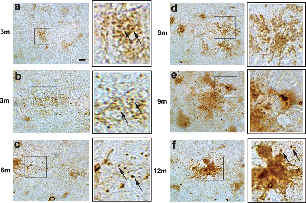
Fig. 7.
Immunohistochemical analysis of the distribution of pE(3)Aβ with the D129 antibody in young and old mThy1-AβPP tg mice. Immunohistochemistry on vibratome sections from the frontal cortex of mThy1-AβPP tg mice from 3 mo to 12 mo of age. a, b) Representative micrographs of D129 immunoreactivity in the frontal cortex of 3 m mThy1-AβPP tg mice, insets at higher magnification. c) Representative micrograph of D129 immunoreactivity in the frontal cortex of 6 m mThy1-AβPP tg mouse, inset at higher magnification. d, e) Representative micrographs of D129 immunoreactivity in the frontal cortex of 9 mo mThy1-AβPP tg mice, insets at higher magnification. f) Representative micrograph of D129 immunoreactivity in the frontal cortex of 12 mo mThy1-AβPP tg mouse, inset at higher magnification. Scale bar = 10 µM.

