Fig. 8.
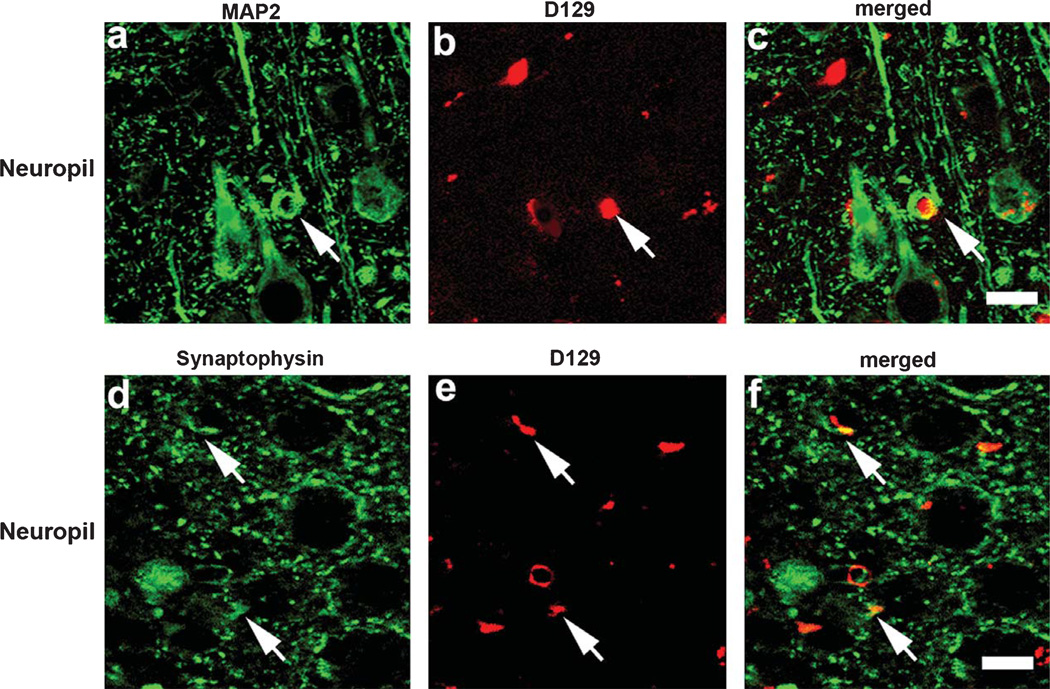
Double immunolabeling studies for pE(3)Aβ and dendritic and synaptic markers co-localization in mThy1-AβPP tg mice. Co-localization studies were performed with the D129 antibody and the dentritic marker MAP2 or the synaptic marker synaptophysin in the frontal cortex of 3 m mThy1-AβPP tg mice. a–c) Representative confocal images depicting co-localization between MAP2 and D129 immunoreactivity in the frontal cortex of 3 mo mThy1-AβPP tg mice. d–f) Representative confocal images depicting co-localization between synaptophysin and D129 immunoreactivity in the frontal cortex of 3 mo mThy1-AβPP tg mice. Arrows indicate areas of signal co-localization. Scale bar = 30 µM.

