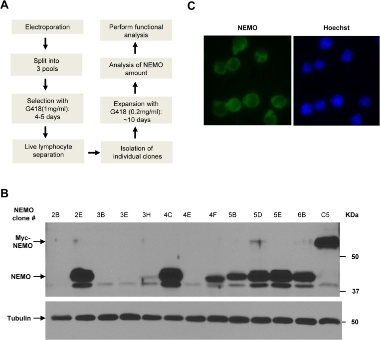
Fig 3. Generation of 1.3E2 clones expressing different amounts of NEMO protein.
(A) Work flowchart to generate NEMO reconstituted 1.3E2 cell clones. (B) An example of Western blots with different NEMO stable clones. Protein extracts from different 1.3E2 clones were separated on SDS-PAGE gel and immunoblotted with NEMO antibody. Tubulin was used as a loading control. (C) An example of immunofluorescence analysis with NEMO antibody (Green) in a NEMO reconstituted 1.3E2 stable clone. Nuclei were visualized by Hoechst staining (Blue).

