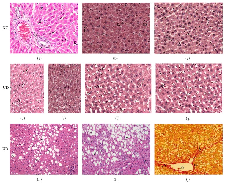
Figure 2.
(a), (b), and (c): histological pattern of liver from nondiabetic control rats (NC) showing normal-appearing of hepatocytes, portal space (PS), sinusoids (arrows), and Kuppfer cells (K) at 6, 14, and 26 weeks of follow-up, respectively. Focal area of mild steatosis (∗) is observed in NC rats sacrificed at 26 weeks. (d) and (e): liver from untreated diabetic rats (UD) sacrificed at 6 weeks showing the onset of sinusoidal enlargement (arrows) and small amount of fatty vacuoles (v), respectively. (f) and (g): liver from UD rats sacrificed at 14 and 26 weeks showing progressive worsening of sinusoidal enlargement (arrows) and liver fatty degeneration (v), respectively. Liver from UD rats at 26 weeks showing in (h) macrovesicular fatty degeneration (v); (i) interlobular mononuclear inflammatory infiltrate consistent with steatohepatitis (∗); (j) periportal (PS) fibrosis (arrows). H&E and red picrosirius (400x).

