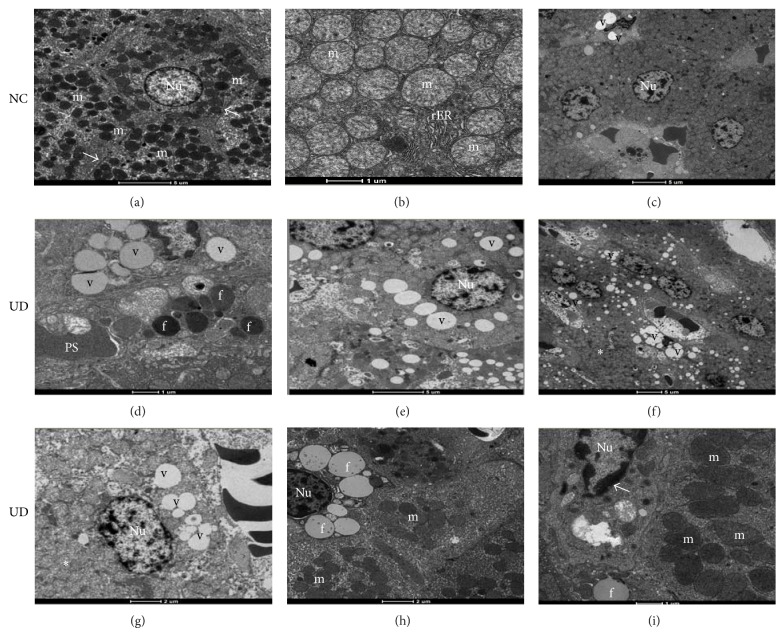
Figure 3.
Electron micrographs of nondiabetic control rats (NC) sacrificed at 6, 14, and 26 weeks, respectively, showing (a) normal-appearing of hepatocytes, nucleus (Nu), mitochondria (m) and Disse's space (arrows); (b) detail of mitochondria (m) and rough endoplasmic reticulum (rER) with their preserved structures; (c) focal area of lipid vacuoles (v) in NC rat sacrificed at 26 weeks (scale bars: (a), (c): 1,900x; (b): 4,800x). (d), (e), and (f): hepatocytes of untreated diabetic rats (UD) sacrificed at 6, 14, and 26 weeks, respectively, showing (d) hepatocyte showing moderate amount of lipid vacuoles (v) and hepatic lipid droplets (f); (e) worsening of liver fatty degeneration (v); (f) poor organization of the organelles within the cytoplasm with disappearance of the Disse's space and rER (∗) with diffuse liver fatty degeneration (v) (scale bars: (d): 4,800x; (e), (f): 1,900x). (g), (h), and (i): hepatocytes of UD rats sacrificed at 26 weeks showing: (g) intense disorganization of cell ultrastructure (∗); (h) scarce amount of mitochondria (m) within the cytoplasm; (i) mitochondria appear larger, less electron-dense with less distinct cristae (m). Note the condensation of nuclear chromatin (arrow) (scale bars: (g), (h), (i): 4,800x).

