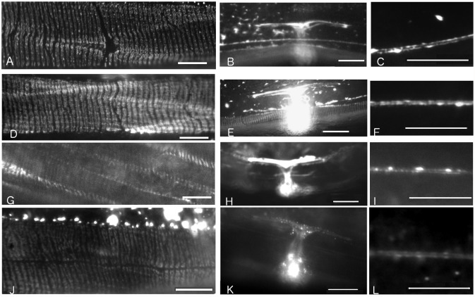
Fig 2. Fluorescence micrographs showing IFA-2::GFP variant localization in live adult transgene carrying lines.
Scale bars for FO and ALM micrographs are 10μm, for uterus micrographs the scale bars are 20 μm. A) Full length IFA-2::GFP fusion protein localizes to epidermal FOs in rescued ifa-2::gfp; ifa-2(nc16) animals. The GFP-dependent fluorescence is detected in thin stripes oriented perpendicular to the long axis of the worm in regions of the epidermis adjacent to muscle. B) Full length IFA-2::GFP fusion protein localizes to uterine seam and to C) ALM in ifa-2::gfp; ifa-2(nc16). D, E, F) Localization of headless IFA-2∆H::GFP protein is indistinguishable from IFA-2::GFP. Genotype shown is ifa-2 ∆H::gfp; ifa-2(nc16) G,H, I) Localization of IFA-2∆T::GFP protein is indistinguishable from IFA-2::GFP. Genotype shown is ifa-2 ∆T::gfp; ifa-2(+). J,K,L) Localization of IFA-2R::GFP protein is indistinguishable from IFA-2::GFP. Genotype shown is ifa-2 R::gfp; ifa-2(+).

