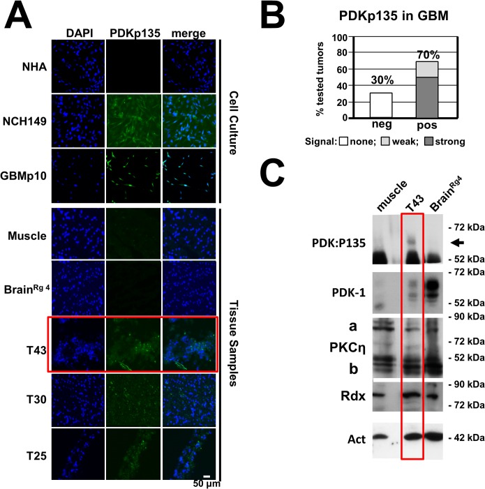
Fig 7. Detection of PDK1phosphoS135 in human cancer tissues.
(A) Cryosections of human brain tumors were analyzed for the presence of PDK1phosphoS135 by immunostaining with specific monoclonal antibodies (PDKp135) and counterstaining with DAPI. Normal human astrocytes, muscle tissue samples and a safety margin of healthy looking brain tissue of tumor #56 were used as negative controls while two glioma-derived cell lines (NCH149, GBM21) served as positive controls. Scale bar, 50 μm. (B) Quantitation of the IF microscopy data (see S6 Fig.) shows that 70% of the analyzed tumor samples (n = 36) contained PDK1phosphoS135-positive cells, 50% with a strong and 20% with a weak signal. (C) The presence of PDK1phosphoS135 in tumor T43 was confirmed by western blot analysis of whole-cell extracts with immunoaffinity-purified phosphospecific antiserum (Rabbit #769). Normal muscle tissue and BrainRg4 were analyzed in parallel for comparison. Total amounts of PDK1, PKCη/Rdx, and actin were also determined.

