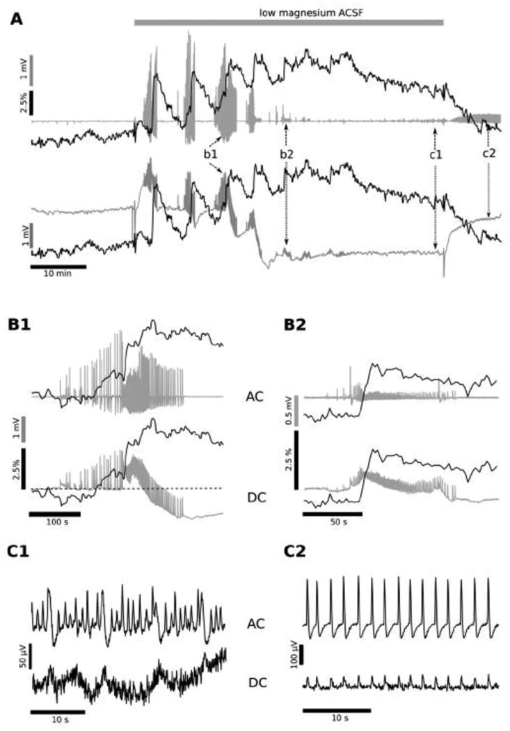
Figure 3. Low Mg2+ condition at P7.

Simultaneous recording of the electrical (local field potential, LFP) and metabolic activity (NADH fluorescence) are shown from an intact hippocampus of the P7 mouse in low magnesium aCSF. Experiments conducted on older, P7 mice showed that the application of low Mg2+ aCSF initially induced the generation of 2-5 large amplitude ILEs (A, b1; detail in B1), then progressively the ILEs decreased in amplitude (A, b2; detail in B2), and transformed into low amplitude (50-400 μV) oscillations, with a frequency of 0.6-1.2 Hz (N=7; A, c1; detail in C1). This low amplitude oscillation, late recurrent depolarization (LRD), persisted even after the switch back to the regular, Mg2+ -containing aCSF (A, c2; detail in C2). Field potential recordings performed in DC mode showed that, similar to experiments in P5 hippocampi, each ILE was accompanied by a baseline negative shift (A, b1, b2: lower trace). The base line behaviour was similar to that observed at P5 during the first 1-2 ILEs (Fig. 1). However, the negative electrical baseline shift grew larger in amplitude (1-3 mV) and irreversible during the last large amplitude ILE (A, b1 lower trace). After this large, negative decrease in the LFP trace the ILE amplitudes decreased (ie, A, b2 as opposed to b1), leading to emergence of LRD behaviour (A, between b2 and c1).
The LFP base line did not completely recover even after the switch to the regular aCSF (between A: c1, c2), continuing 0.5- 1.4 mV more negative that at the beginning of the experiment. The interictal-like events preceding each individual ILE onset were accompanied by a rise in NADH fluorescence (after A, b1), different than the initial, decreased NADH leading up to ILEs in P5 hippocampi (Fig. 1 C1, C2 versus A: c1, c2). The mean amplitude of the NADH fluorescence transient corresponding to the first few ILEs averaged 4.3 ± 1.5% (N=7), but the subsequent ILE transients were smaller in amplitude (B2 vs B1). However, the baseline fluorescence gradually increased over the duration of the exposure to low Mg2+ media, reaching a maximal value of 7.1 ± 2.5% (N=7) at the moment of transition to the LRD generation (A, after b2). Thereafter, the NADH fluorescence remained elevated during the LRD phase (remaining above baseline), and decreased to near the initial value only when the perfusion solution was changed to the regular aCSF (A, after c1). This NADH profile suggests that at P7 the metabolic activity increases not only during each ILE, but also rises prior to ILE occurrence (during the build-up of interictal activity), and continues to increase in the transition to LRD activity, when ILEs are starting to fade. This maintained high NADH level during the transition into LRD activity suggests a profound metabolic demand associated with the ongoing epileptiform activity during this phase.
