Figure 10.
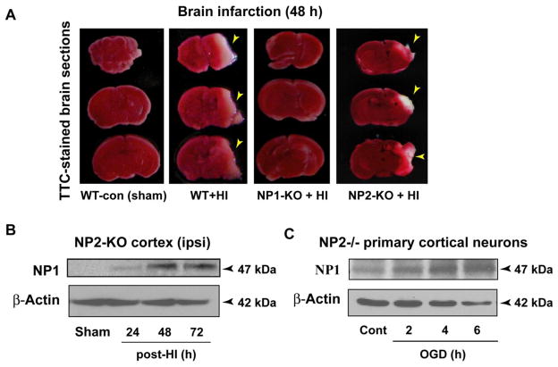
Cerebral HI caused brain infarction in WT and NP2-KO neonatal mice but not in NP1-KO and NP-TKO brains. A) The TTC-staining of brain sections (2 mm) at 48 h post-HI from WT and NP2-KO animals revealed marked infarction (white areas) in the right cerebral cortex and hippocampal CA1 and CA3 areas, which was completely absent in brain sections from NP1-KO and NP-TKO animals under identical HI conditions. Representative images of TTC-stained sections are shown, experiments were repeated twice with n=5 each time. B, C) Western blot analyses of the ipsilateral cortical tissue from NP2-KO brain at different time post-HI showed NP1-specific immunoreactive bands of molecular mass 47 kDa. Similar induction of NP1 was observed in NP2−/− primary cortical neurons following exposure to OGD for different time periods. Representative bands are shown.

