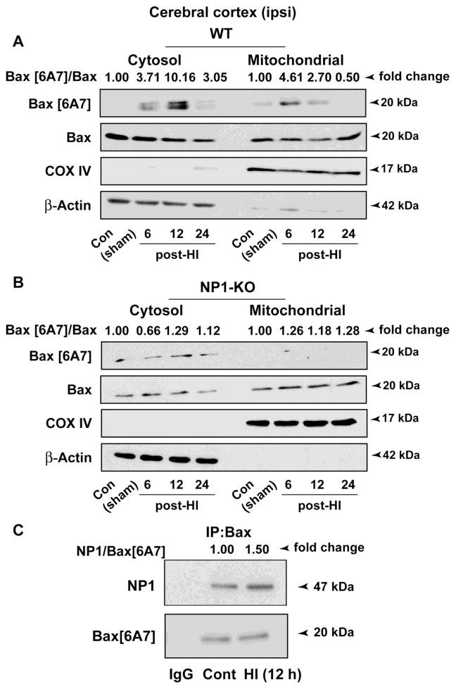
Figure 8.
Effects of HI-induced NP1 on pro-death Bax activation and its translocation to mitochondria. Subcellular fractionation of cortical lysates from WT and NP1-KO brains followed by Western blot analysis showed differential distribution of active Bax [6A7]-specific immunoreactive bands in the cytoplasm and mitochondrial fractions at different time post-HI. A) Increase in active Bax [6A7] protein levels was apparent in mitochondrial fraction at 6 and 12 h post-HI. In contrast, the Bax [6A7] specific bands were absent or remained unchanged in the absence of NP1 expression in NP1-KO brains similar to that observed in respective controls (B). COXIV and actin were used as control for purity of the mitochondrial and cytosolic fraction, respectively, which also served as loading controls. Values are shown as ratio of Bax[6A7]/Bax and expressed as fold change over sham controls from four separate experiments (n=4). Representative bands are shown. C) Interactions of NP1 with the active Bax. SDS-PAGE and immunoblotting of Bax[6A7] immunoprecipitates with NP1- and Bax[6A7]-specific antibodies revealed HI-induced increase of NP1 co-precipitation (1.5-fold) relative to sham control. IgG immunoprecipitates showed no evidence of NP1- and Bax-specific bands. Representative blots are shown.

