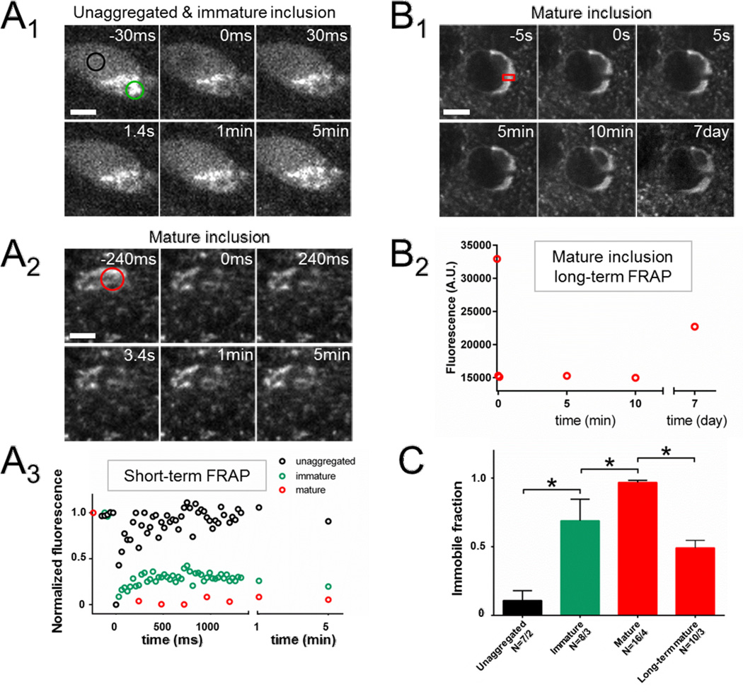
Figure 2. In vivo FRAP demonstrates progressive compaction of and a low molecular turnover rate within Syn-GFP inclusions.
(A) Individual neuron (A1) with an immature inclusion at 3 mpi demonstrates mixed aggregated and unaggregated somatic Syn-GFP staining pattern. Two ROIs, one within (green circle) and one outside (black circle) the aggregated portion are photobleached simultaneously and sequential images shown before and after FRAP. Bleach pulse occurs just before time 0. Scale bar 5 µm. Similar FRAP experiment of the aggregated portion (red circle) of a mature inclusion (A2) at >4 mpi. Scale bar 5 µm. Mean fluorescence intensity (A3) from the three ROIs from the two cells in A1–2 plotted on the same time scale demonstrates complete recovery back to baseline of the homogeneous staining, but large immobile fractions in the immature and mature inclusions.
(B) Individual neuron (B1) with a mature somatic inclusion at 4 mpi and absent unaggregated Syn-GFP staining in the remaining cytoplasm or nucleus. ROI (red rectangle) shows part of inclusion that was photobleached. Scale bar 7 µm. Mean intensity from this ROI over time (B2) shows a large immobile fraction at 5–10 min post-bleach, which recovers substantially at 7 days post-bleach.
(C) Group data comparing measured immobile fractions from unaggregated somatic Syn-GFP, immature and mature inclusions at 5 min post-bleach, and mature inclusions 7 days post-bleach, showing a significant progressive increase in the immobile fraction from immature to mature inclusions as measured at 5 min. At 7 days post-bleach, however, 49% of the previously immobile fraction recovers, demonstrating a slow turnover of protein within inclusions. N = cells/animals.

