
Figure 3. False-positive FDG-PET/CT study.
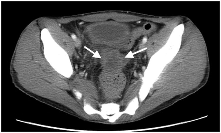
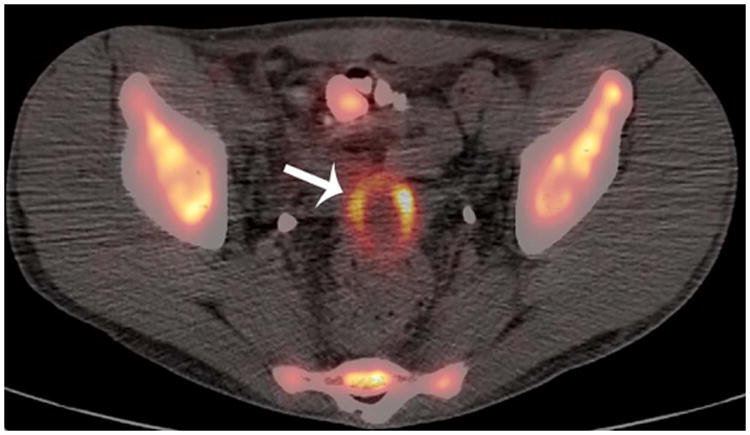
Patient 6 (20-year-old young man treated for Burkitt lymphoma. A) Diagnostic pelvic computerized tomography (CT) shows a residual pelvis mass (arrows). B) Axial positron emission tomography (PET)/CT shows the mass to have intense peripheral fluorodeoxyglucose avidity (arrow). The mass was resected and found to contain necroinflammatory tissue but no viable tumour on histopathological examination.