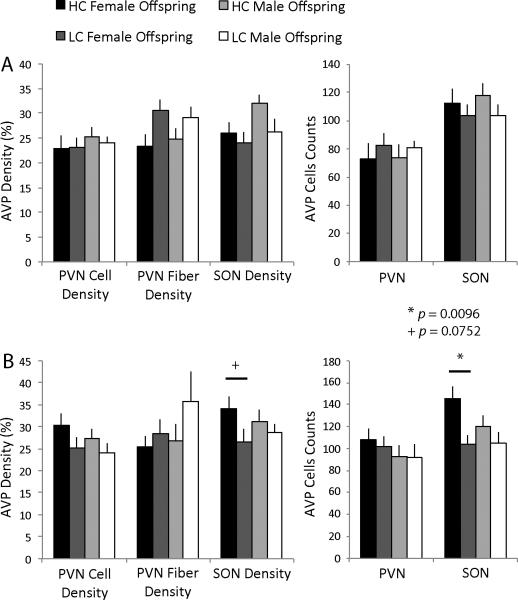
Figure 4.
AVP-ir in the PVN and SON. (A) There were no differences in AVP-ir in the PVN or SON of the hypothalamus following chronic social isolation between HC and LC offspring for either males or females. (B) HC female offspring had a greater number of AVP-labeled cells in the SON compared to LC offspring (F(1,15) = 9.00, p = 0.0096) and tended to also have a greater density of AVP-positive cells and fibers in the SON (F(1,15) = 3.69, p = 0.0752) after an intrasexual aggression test. There were no differences in males in AVP-ir following varying early care.

