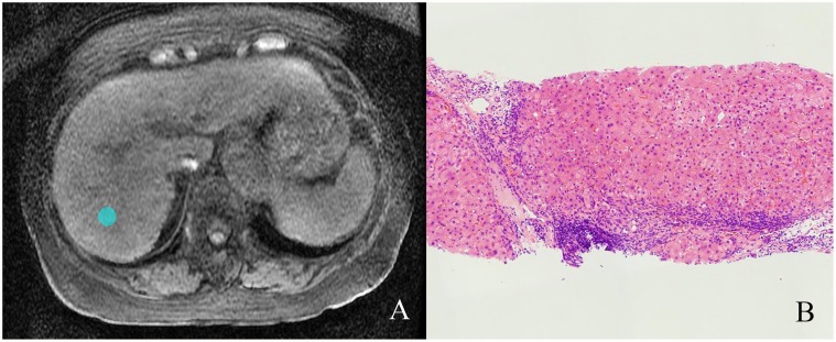
Fig 1. Illustration of region of interest (ROI).
(A) One 20-pixel diameter ROI was placed in the axial T1W images (3.6/1.6; flip angle, 12°) at the level of the porta hepatis in a 76-year-old woman with chronic hepatitis C. (B) Liver biopsy specimen showed stage 4 hepatic fibrosis and grade 2 necroinflammatory activity. (Hematoxylin-eosin stain; original magnification, ×50.)

