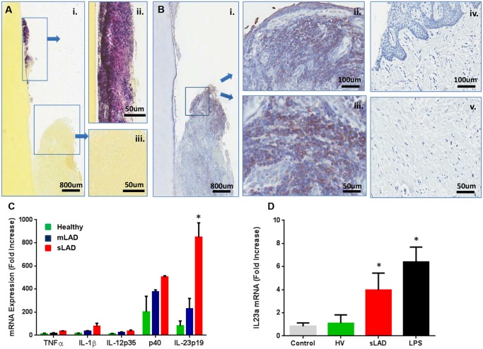
Fig 5. Immunostimulatory potential of LAD- associated dental plaque.
(A) Gram staining (Brown and Brenn method) of extracted tooth and adjacent soft tissues: (i) Gram positive (violet) and negative (pink) staining on the root surface (ii) and surrounding tissues (iii)(scale bars shown). Representative of 3 LAD patients with tooth extractions. (B) Immunohistochemistry for bacterial lipopolysaccharide (LPS) on extracted LAD tooth (i) and surrounding tissues (ii, iii) as well as in healthy gingiva (iv, v). Positive staining is brown and indicated by arrows (scale bars shown). Representative of 3 LAD patients. (C) Exposure of human macrophages to LAD microbial plaque. Human macrophages (at a concentration of 3x10⁶/ml) were left untreated (medium control) or treated with standardized amounts of inactivated subgingival plaque (equivalent of 1x10⁶ 16S rRNA gene copy number) from healthy, mLAD and LAD donors (n = 3, each) for 4 hours and processed for RNA to evaluate cytokine gene transcription. Results are shown as fold induction of mRNA expression relative to the untreated control, p<0.05 between health and LAD. (D) In vivo inoculation of murine oral mucosa with inactivated subgingival plaque from healthy volunteer (HV) and sLAD donors or purified LPS (2 μg/μl). Mice received oral injections with HV or sLAD plaque or LPS and oral tissues were harvested at 4h for evaluation IL23a at the mRNA level (n = 3). Results are shown as fold increase over untreated control, * = p<0.05, significant increase over treatment with HV plaque.

