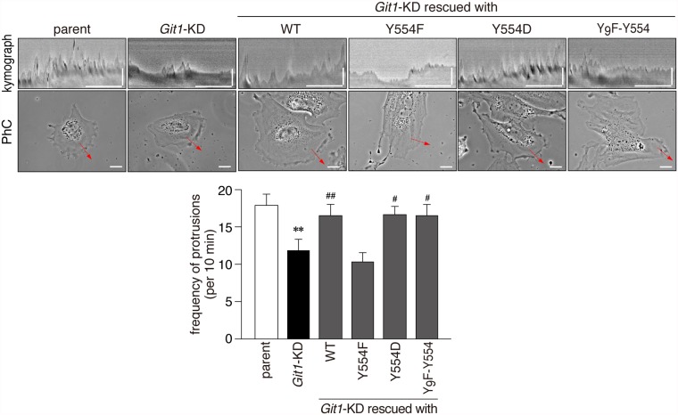
Fig 9. The phosphorylation-defective Tyr-554 mutant of Git1 failed to restore impaired lamellipodial protrusion activity in A7r5 cells by Git1-knockdown.
Representative kymographs (upper pictures; scale bars, 10 min on the x-axis and 10 μm on the y-axis) obtained from phase-contrast photographs (lower pictures; scale bars, 20 μm) of parental cells, Git1-KD cells, and Git1-KD cells expressing various mCherry-fused Git1 proteins on fibronectin-coated dishes. Before beginning the recording, the expression of mCherry-fused proteins was verified in the rescue experiments by a short exposure to UV to avoid possible cell damage. The lower graphs show the frequency of protrusions. Data are the mean ± S.E. (error bars; n = 10 to 11 each). **, P < 0.01 significantly different from parental cells; #, P < 0.05 or ##, P < 0.01 significantly different from Git1-KD cells by ANOVA and Scheffé’s post hoc tests.

