
Figure 1.
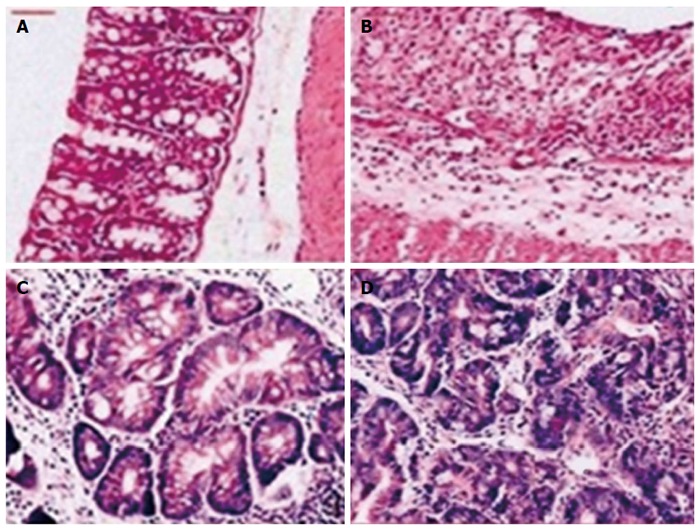
Hematoxylin and eosin staining of colons obtained from rats in each group. A: Normal colonic histology (× 20); B: Colonic histology from a rat with ulcerative colitis (× 20); C: Dysplastic lesion in a rate with colorectal cancer (× 200); D: Cancer lesion in a rat with colorectal cancer (× 200).