
Figure 2.
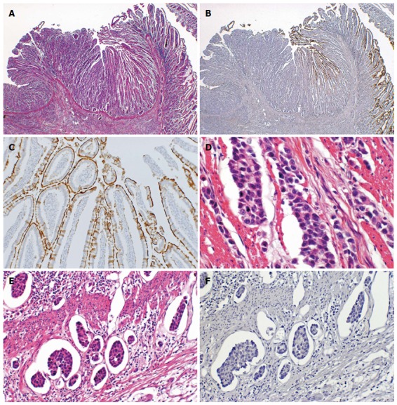
Representative CD10-negative small intestinal adenocarcinoma case. A: Normal small intestinal mucosa was observed on the right side, whereas the adenocarcinoma was observed on the left side (HE stain, × 12.5); B: Normal mucosa was positive for CD10, but adenocarcinoma was negative for CD10 (CD10, × 12.5); C: The brush border of the normal mucosa was positive for CD10 (CD10, × 100); D: This SIA case showed a poorly differentiated adenocarcinoma (HE satin, × 200); E: Lymphatic permeation was frequently seen (HE stain, × 200); F: The carcinoma cells were negative for CD10 (CD10, × 200).