
Figure 1.
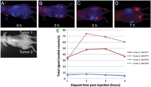
Specific targeting of tumor with fluorocoxib A and its imaging timecourse in the BCC allograft model. Time-lapse in vivo fluorescence imaging of BCC allograft mice after co-injection of Fluorocoxib A (LM-4777) and LiCor800-equivalent (LM-5516) probes. Time sequence is (A) 0 hours (B) 3 hours (C) 5 hours (D) 7 hours post-injection, where the allograft mouse is oriented as in the brightfield image below (A). The red signal represents LM-4777 and the blue signal represents LM-5516. (E) Total unmixed fluorescence signal of tumor 1 and 2 at each time point for LM-4777 and LM-5516.