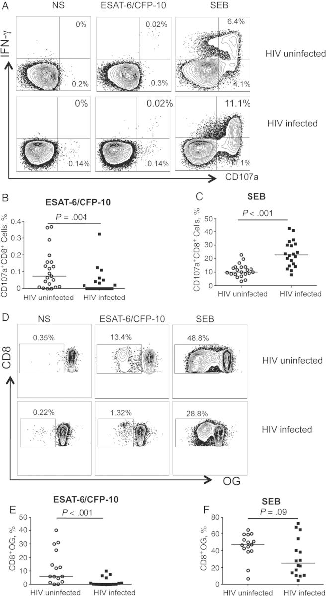
Figure 2.

Impaired degranulation and proliferation of Mycobacterium tuberculosis–specific CD8+ T cells in human immunodeficiency virus (HIV)-infected compared with HIV-uninfected individuals with latent M. tuberculosis infection. A, Degranulation activity in M. tuberculosis–specific CD8+ T cells was assessed by expression of cell surface CD107a. Representative flow cytometry plots for an HIV-uninfected and an HIV-infected individual with latent tuberculosis demonstrate CD107a expression by CD8+ T cells in nonstimulated (NS) cells (left), on stimulation with ESAT-6/CFP-10 peptide pools (middle) and Staphylococcus enterotoxin B (SEB) (right). B, C, Summary data comparing frequencies of CD107a+CD8+ T cells between HIV-uninfected (n = 21) and HIV-infected (n = 19) groups on stimulation with ESAT-6/CFP-10 (B) or SEB (C). Horizontal lines indicate median frequency of CD107a+CD8+ T cells. D, Proliferative capacity of M. tuberculosis–specific CD8+ T cells was measured after a 6-day stimulation of freshly isolated peripheral blood mononuclear cells with CFP-10 and ESAT-6 peptide pools. Representative flow cytometry plots for an HIV-uninfected and an HIV-infected individual with latent tuberculosis demonstrate proliferation of CD8+ T cells on stimulation with ESAT-6/CFP-10 (middle) or SEB (right). The cells shown are gated on VIVIDloCD3+CD8+ lymphocytes. The percentage on each plot indicates the percentage of proliferating CD8+ T cells after subtraction of background proliferation in the uninfected control condition. E, F, Summary data comparing frequencies of proliferating CD8+ T cells between HIV-uninfected (n = 15) and HIV-infected (n = 16) individuals on stimulation with ESAT-6/CFP-10 (E) or SEB (F). Horizontal lines indicate median frequency of proliferating CD8+ T cells. Differences in the frequencies of CD107a+CD8+ T cells or proliferating CD8+ T cells between HIV-uninfected and HIV-infected groups were assessed using the Mann–Whitney test; differences were considered statistically significant at P < .05 (2 tailed). Abbreviations: IFN, interferon; OG, Oregon Green; VIVID, violet dead cell stain.
