
Figure 5.
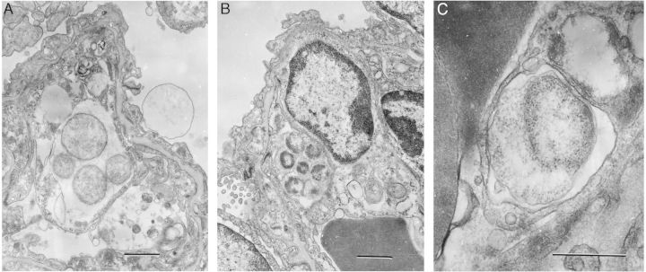
Ultrastructure of Ehrlichia muris–like agent in infected lung during lethal infection. A, A morula-containing reticulate cells (RC) in the cytoplasm of a lung cell. The bar denotes 1 µm. B, A morula with dense-core cells and intramorular fibrillar material in the cytoplasm of an endothelial cell. The bar denotes 1 µm. C, A small morula with an RC with expanded periplasmic space in the cytoplasm of an endothelial cell. Dark masses on the left represent parts of erythrocytes in a capillary lumen. The bar denotes 0.5 µm.