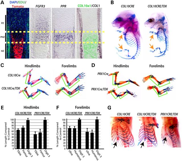
Figure 1.
Mutant activated FGFR3 activity in prehypertrophic chondrocytes of the growth plate is responsible for the majority of the long bone growth defect in TDII. (A) Location of CRE-sensitive Rosa-Tomato reporter activity in COL10CRE mice at E14.5 in relation to cell proliferation (EDU incorporation) and expression of FGFR3, PPR, and COL1 RNA and COL10a1 protein. The yellow dashed lines demarcate the prehypertrophic zone (PHZ). The approximate locations of the proliferative and hypertrophic zones (PZ and HZ) are indicated. (B) Representative examples of Alcian blue- and Alizarin red-stained COL10CRE and COL10CRE;TDII littermates at E15.5. (C) Alcian blue- and Alizarin red-stained hindlimbs and forelimbs from COL10CRE and COL10CRE;TDII littermates and (D) PRX1CRE and PRX1CRE-TDII littermates at E18.5. Green brackets demarcate the long bones. (E and F) The mean percentage of the length of COL10CRE;TDII (E) and PRX1CRE;TDII (F) hindlimb and forelimb long bones and digit three compared with littermate control long bones and autopods at E18.5. Standard deviations are shown (N = 6 for COL10CRE;TDII and COL10CRE littermates and N = 5 for PRX1CRE;TDII and PRX1CRE littermates). (G) Views of Alcian blue/Alizarin red-stained sternum and rib cages of E18.5 COL10CRE and COL10CRE;TDII littermates and a representative PRX1CRE;TDII embryo.

