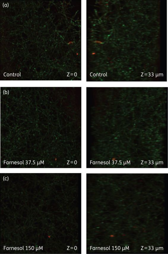
Figure 4.

Effect of farnesol on C. albicans biofilms. (a) Untreated biofilm. (b) Biofilm exposed to farnesol at 37.5 μM. (c) Biofilm exposed to farnesol at 150 μM. Concanavalin A-Alexa Fluor 647 conjugate (green stain highlighting the Candida cell walls) and FUN-1 (yellow-orange stain highlighting non-viable cells) were used to directly visualize the effects of antifungal agents on biofilms. Images are single optical sections. The first image in each row depicts the upper layer of the biofilm, while the second panel illustrates a level near the bottom of the biofilm. This figure appears in colour in the online version of JAC and in black and white in the print version of JAC.
