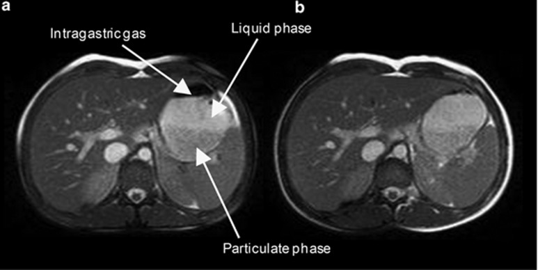
Figure 1.
Representative example MRI images of the stomach of a volunteer taken at the first time point after eating (a) the high-carbohydrate and (b) the high-fat meal on two separate occasions. In the images of the stomach the less intense lower portion belongs to the particulate phase of the meal while the brighter upper phase belongs to a fluid layer. The black part on the top of the stomach is a pocket of intra-gastric gas.

