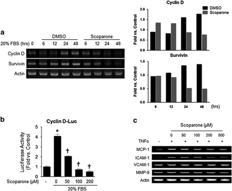
Figure 2.
Transcriptional regulation by scoparone in vascular smooth muscle cells (VSMCs) or HepG2 cells. (a) RT–PCR. The mRNA expression levels of cyclin D, survivin D and actin as a control are shown in representative agarose gels. Total RNA was isolated from VSMCs after incubation with 20% FBS±scoparone (200 μM) for different durations (0–48 h). (b) Promoter activity assay by luciferase reporter assay in HepG2 cells. Cells were incubated with 20% FBS±scoparone (0–200 μM) for 24 h after cyclin D promoter region-luciferase transfection. Relative luciferase activities were determined and normalized according to the activities of the control cells. (c) Semi-quantitative PCR was performed to assess the expression of chemokines and cell adhesion molecules, such as MCP-1, ICAM-1, VCAM-1, MMP-9 and actin as a control. VSMCs were treated with TNF-α (10 ng ml−1) under different scoparone concentrations (0–500 μM) for 24 h. *P<0.05 vs control and †P<0.05 vs 20% FBS (treated or TNF-α only).

