Figure 5.
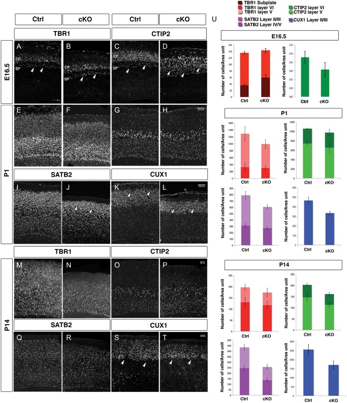
Cortical layering is altered in Arx cKO brains. Immunohistochemistry for molecular markers identifying deeper cortical layers at E16.5 and both deeper and upper cortical layers at P1 and P14, in control and Arx cKO cortices (A–T). (A and B) TBR1+ cells are slightly decreased in layer V–VI neurons and increased in subplate in E16.5 Arx cKO cortices with respect to the controls (aarowheads). (C and D) CTIP2 immunolabeling shows the same alterations observed with TBR1 although more prominently (arrowheads). (E and F) At P1, similar numbers of TBR1+ cells in Arx cKO cortices with respect to control are detected as well as a similar number of CTIP2+ cells in both layers V and VI (G and H). (I and J) SATB2 is decreased in upper layer neurons (II, III) in the Arx cKO cortices with respect to the controls (arrowheads). (K and L) Similarly, staining for CUX1 (layers II–IV) shows a diminution of upper cortical layer neurons in Arx cKO cortices with respect to controls (arrowheads). (M–P) At P14, there are still similar numbers of TBR1+ and CTIP2+ cells in the Arx cKO cortices when compared with controls but still a decrease in SATB2+ cells in layers II/III (Q, R). Similarly, there are also still fewer CUX1+ cells in the Arx cKO cortices (S and T). (U) Quantifications of TBR1+ and CTIP2+ cells in layers VI and V and of SATB2+ and CUX1+ at E16.5, P1, and P14 in both control and Arx cKO cortices. TBR1+ and CTIP2+ cells at P14. Cells positive for each molecular marker were counted 25% of the cortex in P1 and in a 200-μm bin in P14, both from the ventricular to the pial surface in both control and Arx cKO cortices.

