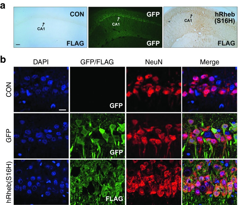
Figure 1.
Transduction of hippocampal neurons with AAV-GFP and AAV-hRheb(S16H) in normal adult SD rats. (a) Animals received a unilateral injection of AAV-GFP or AAV-hRheb(S16) in the hippocampus and were sacrificed 4 weeks later. AAV-hRheb(S16H)-injected brain sections (30 μm) were processed for immunostaining with antibodies against FLAG. The expression of GFP and FLAG (brown reaction product) is observed in the hippocampus for each viral injection, but no expression of FLAG is seen in the noninjected control side (CON). Bar = 100 μm. (b) Immunofluorescence triple labeling for DAPI (blue), GFP (green), and NeuN (red), or DAPI, FLAG (green), and NeuN shows that transgene expression is identifiable within neurons in the CA1 of hippocampus. Bar = 20μm. All of the pictures show the representative coronal sections following each immunostaining (n = 3, each group).

