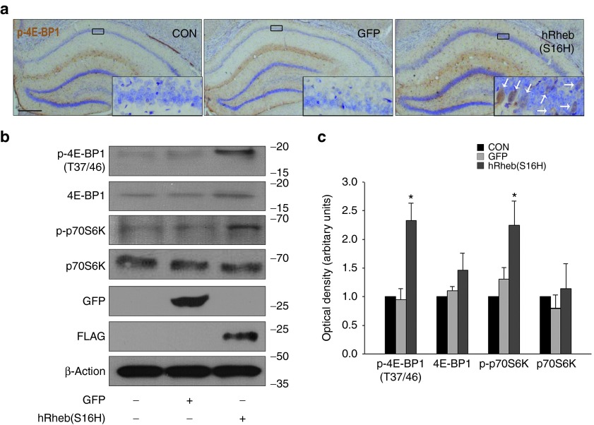
Figure 2.
hRheb(S16H) expression activates mTORC1 in the hippocampus. (a) Brain sections were stained with anti-phospho-4E-BP1, a mTORC1 substrate, at 4 weeks postinjection of viral vectors. Immunoperoxidase staining for p-4E-BP1 (with thionin counterstain) shows that brown reaction products are clearly observed in the neurons of the hRheb(S16H)-treated group, compared to a modest level in the vehicle-treated group. Bar = 500 μm. Insets show magnified photomicrographs of the area in the CA1 layer. An example of neuronal p-4E-BP1 staining (white arrows) is shown in the inset. All pictures show the representative coronal section of each group (n = 3, each group). (b) Western blot analysis of p-4E-BP1, 4E-BP1, p-p70S6K, and p70S6K expression at 4 weeks after intrahippocampal injection of AAV-GFP and AAV-hRheb(S16H). Successful transduction of the hippocampus was confirmed in each case by western blot analysis of GFP and FLAG expressions. (c) The histogram results show the results of a quantitative analysis based on the density of the p-4E-BP1, 4E-BP1, p-p70S6K, and p70S6K bands normalized with the β-actin band for each sample. All values represent the mean ± SEM of four pooled samples for each group. *P < 0.01, significantly different from contralateral control side (CON) and AAV-GFP (one-way analysis of variance and Student–Newman–Keuls analysis).

