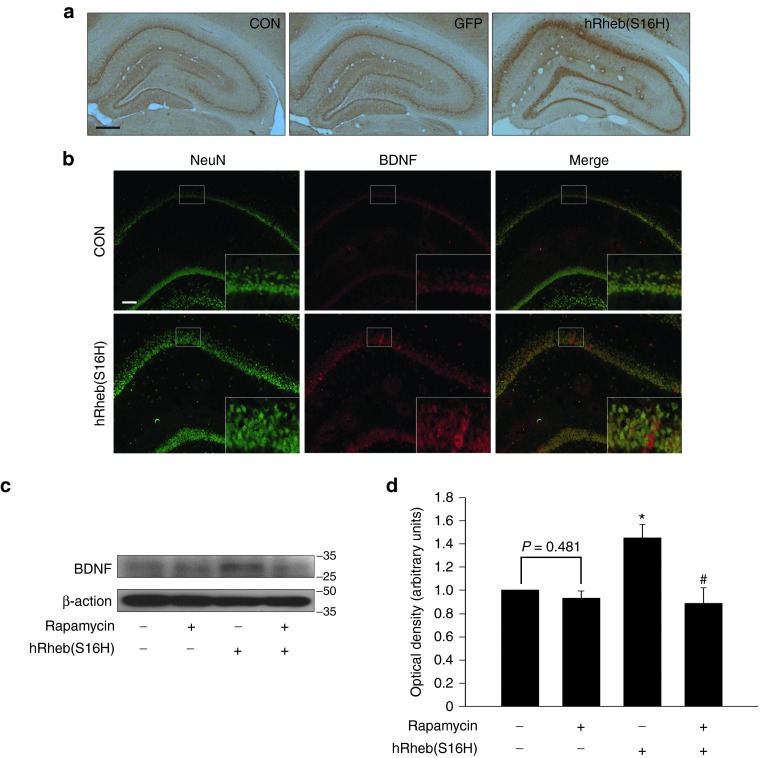
Figure 4.
hRheb(S16H) increases BDNF expression in hippocampal neurons. (a) Immunoperoxidase staining for BDNF was performed at 4 weeks postinjection of AAV-hRheb(S16H). Both the contralateral control side (CON) and the AAV-GFP-injected side (GFP) show similar levels of BDNF in the hippocampus. However, the AAV-hRheb(S16H)-injected side (hRheb(S16H)) shows an increase in the level of BDNF in the hippocampus. Bar = 500 μm. (b) Immunofluorescence double staining for NeuN (green) and BDNF (red) shows that the hRheb(S16H)-increased expression of BDNF is identifiable within hippocampal neurons. Bar = 100 μm. Insets are higher magnifications of each CA1 layer. All pictures in a and b show representative coronal sections following each immunostaining (n = 3, each group). (c) Western blot analysis of BDNF in the hippocampus. Rats were intraperitoneally injected with rapamycin (5 mg/kg), starting 3 weeks after AAV-hRheb(S16H) injection and continued daily injection until 4 weeks postinjection, and then they were sacrificed for western blot analysis. The results show that treatment with 5 mg/kg rapamycin alone causes no alteration to the basic level of BDNF compared to the intact control (first band). However, its treatment attenuated the level of hRheb(S16H)-increased BDNF. (d) All values of the optical density of each band represent the mean ± SEM of six pooled samples in each group. *P = 0.002 and #P < 0.001, significantly different from CON and hRheb(S16H) alone, respectively (one-way analysis of variance and Student–Newman–Keuls analysis).

